Prokaryotic viruses have evolved various mechanisms to transport their genomes across bacterial cell walls, barriers that can contain two lipid bilayers and a peptidoglycan layer. Many bacteriophages utilize a tail to perform this function, whereas tail-less phages rely on host organelles, such as plasmid-encoded receptor complexes and pili. However, phiX174-like bacterial viruses do not fall into either of these well-defined infection paradigms. To penetrate cells, coliphage phiX174 utilizes 10 to12 copies of its pilot protein H that is monomeric during the entire particle assembly pathway. Lei Sun from the Rossmann lab at Purdue University and Ben Fane at the University of Arizona have shown that the central domain of the H protein assembles into 170-Å long tubes using data collected at GM/CA @ APS. The tube is constructed of 10 alpha-helices with their N-terminal region arrayed in a right-handed super-helix coiled coil and their C-terminal region arrayed in a left-handed super-helix coiled-coil. They have used genetic and biochemical data to demonstrate that the tube is essential for infectivity but does not affect in vivo virus assembly. As there is no evidence for the presence of the tubes in mature virions by cryo electron microscopy, the monomeric components do not assemble until the virus recognizes its host. They have also shown that these tubes span the periplasmic space to transport the viral genome into the host's cytoplasm. The 23-Å wide central channel of the H protein tubes is lined with amide and guanidinium side chains. Bioinformatic analyses indicate that this may be a general property of viral DNA conduits and is likely to be critical for efficient genome translocation into the host.
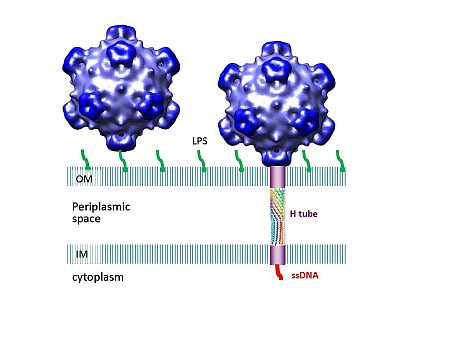 |
Figure: PhiX174 forms a tail for DNA transport during infection. One of the spikes of the virus recognizes a lipopolysaccharide (LPS) molecule in the outer membrane (OM) of the E. coli cell wall. After attachment, the virus extrudes H proteins and a tail forms for DNA penetration. The tail tube is shown crossing the periplasmic space, lodged in between the outer and inner membrane (IM). |
Citation: Sun L, Young LN, Zhang X, Boudko SP, Fokine A, Zbornik E, Roznowski AP, Molineux IJ, Rossmann MG, Fane BA. Icosahedral bacteriophage [phi]X174 forms a tail for DNA transport during infection. Nature. 2014 January 16; 505: 432-435. doi: 10.1038/nature12816
